 es
es en
en fr
fr it
it ru
ru ar
ar zh-hans
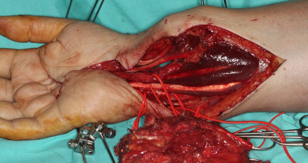
The patient, a young male, has a recurrent tumor in the area of the wrist likely to eventually become malignant after appearing for a third time.
After the last diagnosis, an amputation is recommended due to disabling pains and the risk of metastasis.
The problem
The analysed picture presents characteristics that make malignancy and the appearance of a metastatic process likely. The patient visits the clinic of Dr. Piñal with the loss of the hand as the most probable outcome, once the tumor mass already extends through the metacarpals and occupies the area of the wrist almost in its entirety.
The objectives
The main goal of Dr. Piñal’s surgical unit is to preserve the limb, safeguarding its functional characteristics, as well as to eliminate pain and to mitigate the final aesthetic impact.

The plan
Dr. Piñal designs an intervention that starts from the resection (surgical removal) of the tumor with a safety margin that minimizes the risk of recurrence, acting on an area of 10 cm from the metacarpals to radius and cubit.

The right image shows the removal of all affected tissues and tendons, leaving only the viable and essential elements for the subsequent microsurgical reconstruction: the median nerve, which gives sensitivity to the fingers, the ulnar nerve and the flexor tendons.

After the removal of about 400 grams of tumor mass, the challenge lies in the osseous and functional restoration of the affected area, which, moreover, must be given back blood flow. To do this, Dr. Piñal uses an autologous microsurgical graft of vascularized fibula (the patient himself acts as a donor), split in three to form a delta shaped structure that covers the defect.


The procedure of insertion and nervous and vascular microsurgical reconnection lasts for eleven hours.
The fibula plays an accessory role in the general ‘load’ of the lower limb, approximately 10% of that of the tibia, whereby the effects on the patient are negligible in relation to the benefit obtained.
The results
After a radiation treatment to ensure a complete disappearance of the tumor, the patient stops feeling pain and recovers function, with a limited aesthetic condition, as can be seen.

