 es
es en
en fr
fr it
it ru
ru ar
ar zh-hans
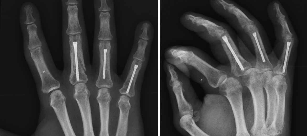
The patient, a middle-aged male, presents severe multifragmentary crushing fractures of the proximal phalanges in the third, fourth and fifth fingers of his left hand. Dr. Piñal choose his technique of insertion of cannulated screws to facilitate the osteosynthesis.

The problem
Multifragmentary or comminuted fingers fractures present the additional difficulty of the bone’s break into various fragments. A conventional approach by means of bone fixation with Kirschner needles lengthens the duration of the procedure and prevents the patient from mobilizing until 3-4 weeks after surgery.
Axial tomography (CT) images allow the preoperative picture of multifragmentary fractures of the patient’s proximal phalanges to be observed.
During this waiting period complications such as the adhesion or ‘gluing’ of the tendons to the bone may arise, which causes loss of function in the affected area after fracture recovery.

Together with bone damage, the trauma suffered causes the patient serious injuries, prior to his arrival at the clinic of Dr. Piñal.

The objectives
The fundamental goals of Dr. Piñal and his surgical unit are to achieve an osteosynthesis, that is to say, the reduction-healing of the fractures, that facilitates an immediate postoperative mobilization and that reduces the time of recovery.
In turn, the elimination of any subsequent discomfort and the minimizing the aesthetic impact of the procedure are pursued.

The plan
In this case, Dr. Piñal uses his minimally invasive fixation technique for phalangeal and metacarpal fractures, by means of intramedullary insertion of headless cannulated screws, between 3 and 4 mm in diameter depending on the type of injury. It is an ambulatory surgery modality, that is, the patient is discharged after the intervention without needing a hospital stay.
This pioneering development of the Spanish surgeon was first described in his article ‘Minimally Invasive Fixation of Fractures of the Phalanges and Metacarpals with Intramedullary Cannulated Headless Compression Screws’, published by the journal of the American Society for Surgery of the Hand in April 2015.

As the first key step in the surgery, Dr. Piñal introduces a metal guide into each fractured finger, which adjusts the reduction and allows each cannulated screw to be inserted and slid into place. The appropriate length of such screws is pre-established through axial tomography (CT).

However, in comminuted fractures, such as those of the patient, compression without adequate bone support would be ineffective, so an axial strutting is performed, which causes the cannulated screw to act as a girder on the base of each phalange affected

In the images, development of the technique of insertion of cannulated screws by Dr. Piñal in a case of middle phalanx fracture.
After just an hour in the operating room, well below the time required in other techniques, the patient is allowed to start moving his fingers immediately.
The results
As the inferior set of images allows to see, the surgery is a success, with visible effects already in the first two weeks after the intervention.
In the video: functional and aesthetic recovery of the patient in various moments of the period after the intervention of Dr. Piñal: week 1, 2, 3 and state at three months.
Additional info:
- ‘The insertion of cannulated screws by Dr. Piñal shortens the recovery time in finger fractures’.
- The second edition of TVE’s Telediario (Spanish public broadcaster night bulletin) shows the technique of cannulated screws used by Dr. Del Piñal in the treatment of finger fractures: