 es
es en
en fr
fr it
it ru
ru ar
ar zh-hans
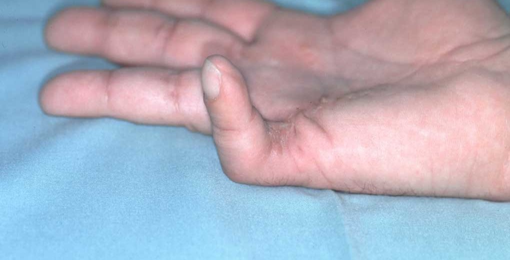
Ci-dessous vous pouvez lire une traduction de traducteur humain en anglais. Vous avez également accès à l’original espagnol en cliquant sur le drapeau dans le coin supérieur droit. Ce lien vous donne accès à une version de traduction automatique de Google en français: https://bit.ly/3ijbxrs
Vascularized neurocutaneous free flaps in Dupuytren cases may be the best option when the affected hand bed is poor, the defect is long or there is a loss of concomitant tissue, variables typical of cases with previous surgery. This is the starting situation of the patient under analysis, a 53-year-old male with an advanced Dupuytren’s picture on the fifth finger of his right hand, contractured in flexion towards the palm.

Dupuytren’s contracture is more prevalent in men over 60 years, with a ratio of ten to one between male and female patients.
The problem
The patient has a Dupuytren’s contracture in his right little finger, causing functional limitations. In turn, after two unsuccessful surgeries before consulting with Dr Piñal, the damaged area presents a tissue deficit in the palm and palmar face of the fifth finger, to which is added the loss of sensitivity in its ulnar part due to section of the digital nerve in one of the previous interventions.

The goals
The clinical objectives of Dr Piñal and his team are the functional recovery of the hand and the return of sensitivity to the damaged finger, through the microsurgical restoration of the defect.
The plan
Dr Piñal removes fibrous and scar tissue preparing the affected area for the neurocutaneous flap taken from the second toe. This is made up of a cutaneous island with skin and fat, together with two segments of the digital and first intermetatarsal dorsal arteries, the nerve of the donor foot and a subcutaneous vein in order to preserve the free flap irrigation.

During the microsurgery intervention (performed in a single procedure and according to the technique described by Dr. Piñal in The Tibial Second Toe Vascularized Neurocutaneous Free Flap for Major Digital Nerve Defects, J Hand Surg 2007; 32A: 209–217. Copyright © 2007 by the American Society for Surgery of the Hand) the nerve is reconnected in the proximal part of the palm and the little finger pulp, and the arterial segments restore the vascular structure of the area.
The microsurgical connections made in the finger and palm are in a range less than one millimeter in diameter, which is a technical challenge for the surgeon.

The results
The operation allows the patient to recover the flexion-extension movement of the little finger, in addition to returning the lost sensitivity to its ulnar part. Likewise, the procedure designed by Dr. Piñal minimizes the aesthetic impact on the recipient and donor areas.


Related content: