 es
es en
en fr
fr it
it ru
ru ar
ar zh-hans
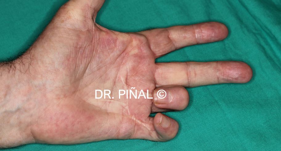
أدناه يمكنك قراءه ترجمه المترجم البشري باللغة الانجليزيه. لديك أيضا الوصول إلى الاسبانيه الاصليه عن طريق النقر علي العلم في الزاوية اليمني العليا. هذا الرابط يتيح لك الوصول إلى نسخه الترجمة اليه من جوجل في العربية
The patient, a 70-year-old man, presents a picture of Dupuytren’s disease in the ring and little fingers of his left hand, which are contractured in flexion (Dupuytren’s contracture) causing a serious functional limitation.

In the development of Dupuytren’s contracture, there is a thickening of the subcutaneous tissue of the palmar fascia that extends to the fingers, and ends up leading to the appearance of bands of fibrous tissue that act as cords or flanges, limiting finger extension.
Dupuytren’s disease is a hereditary pathology without prophylactic treatment, with a higher prevalence in northern Europe, especially in men over 60 years.
In this particular case, the patient underwent two previous surgeries before attending Dr Piñal’s clinic. These procedures generated a tissue deficit in the palm area and the palmar face of the ring and little fingers.

The problem
The patient’s Dupuytren’s contracture is in such an advanced stage that the amputation of both fingers was considered, which Dr. Piñal finds very objectionable as there are other alternatives.
The goals
The fundamental aims of Dr Piñal and his surgical unit in this case are to eliminate the neurocutaneous defect and restore lost function, preserving the sensitivity of the affected area and minimizing the aesthetic impact on both the hand and the donor area of the foot.
The plan

Dr Piñal designs a reconstructive microsurgery plan in a single procedure, which is based on the transfer to the hand of a vascularized free flap of the second toe (according to his own technique described in The Tibial Second Toe Vascularized Neurocutaneous Free Flap for Major Digital Nerve Defects, J Hand Surg 2007; 32A: 209–217. Copyright © 2007 by the American Society for Surgery of the Hand).
The flap is made up of a cutaneous island with skin and fat, accompanied by a segment of digital artery and nerve of the donor foot, and a subcutaneous vein to guarantee the irrigation of the transferred tissue.
After the fibrous bands and scar tissue have been removed, the defect is covered with the neurocutaneous flap: the digital artery of the toe is microsurgically connected to that of the hand, the nerves are also reconnected and, thus, both the enervation and blood flow are restored. The connections made in the proximal part of the palm and the fingertips are within a range of less than a millimeter in diameter (0.5-0.7 mm), which requires a great deal of experience and technical expertise from the surgeon.

The procedure lasts for 4,5 hours and the patient is discharged from hospital after three days.
The results
The intervention carried out by Dr. Piñal recovers a good part of the extension movement of the ring and little fingers, maintaining a sensitivity and appearance of the recipient and donor areas in satisfactory margins for the patient. It should be noted again that after two previous failed surgeries amputation of both fingers had been recommended.

Related content: