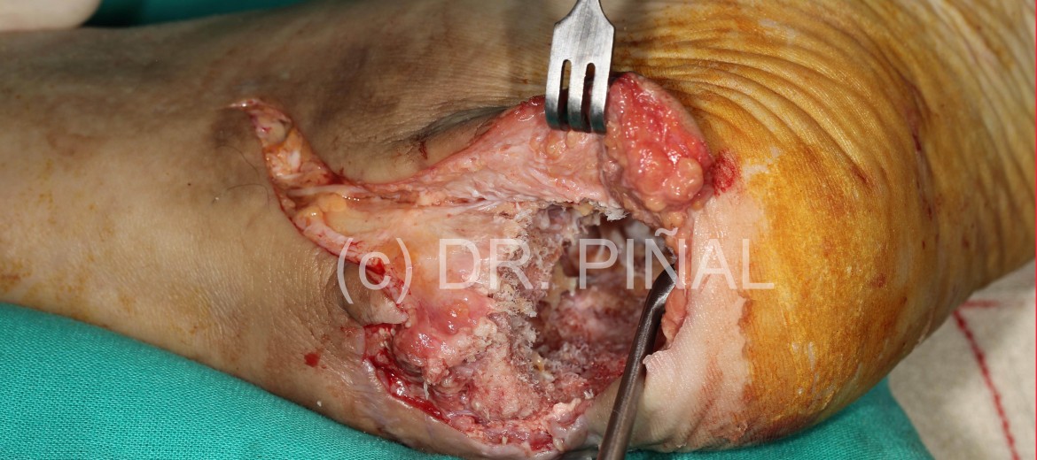
In this case of calcaneal osteomyelitis, the patient, male, 48, comes to the clinic of Dr. Del Piñal after being previously operated in six occasions. After suffering a calcaneal fracture in a fall, the affected area developped an infectious process that was going to be addressed with the amputation of the limb, in absence of other way to stop it.
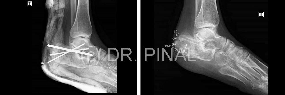
The problem
The persistence of the infection drew a picture of continued pain and restricted mobility, which often ends -as has been pointed out- with the loss of the foot after the the disease becomes chronic.
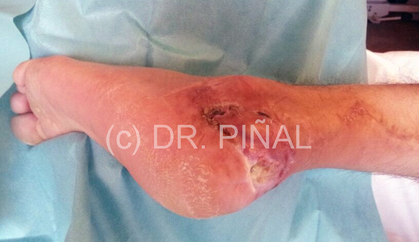
The objectives
The goals of the clinical approach of Dr. Del Piñal in this and other similar cases focus on three main results: eliminate the infection, eradicate pain and preserve the function of the concerned area.
The plan
As an initial step, Dr. Del Piñal makes a calcaneal debridement to clean and clear out the affected area, opening a window through the heel.
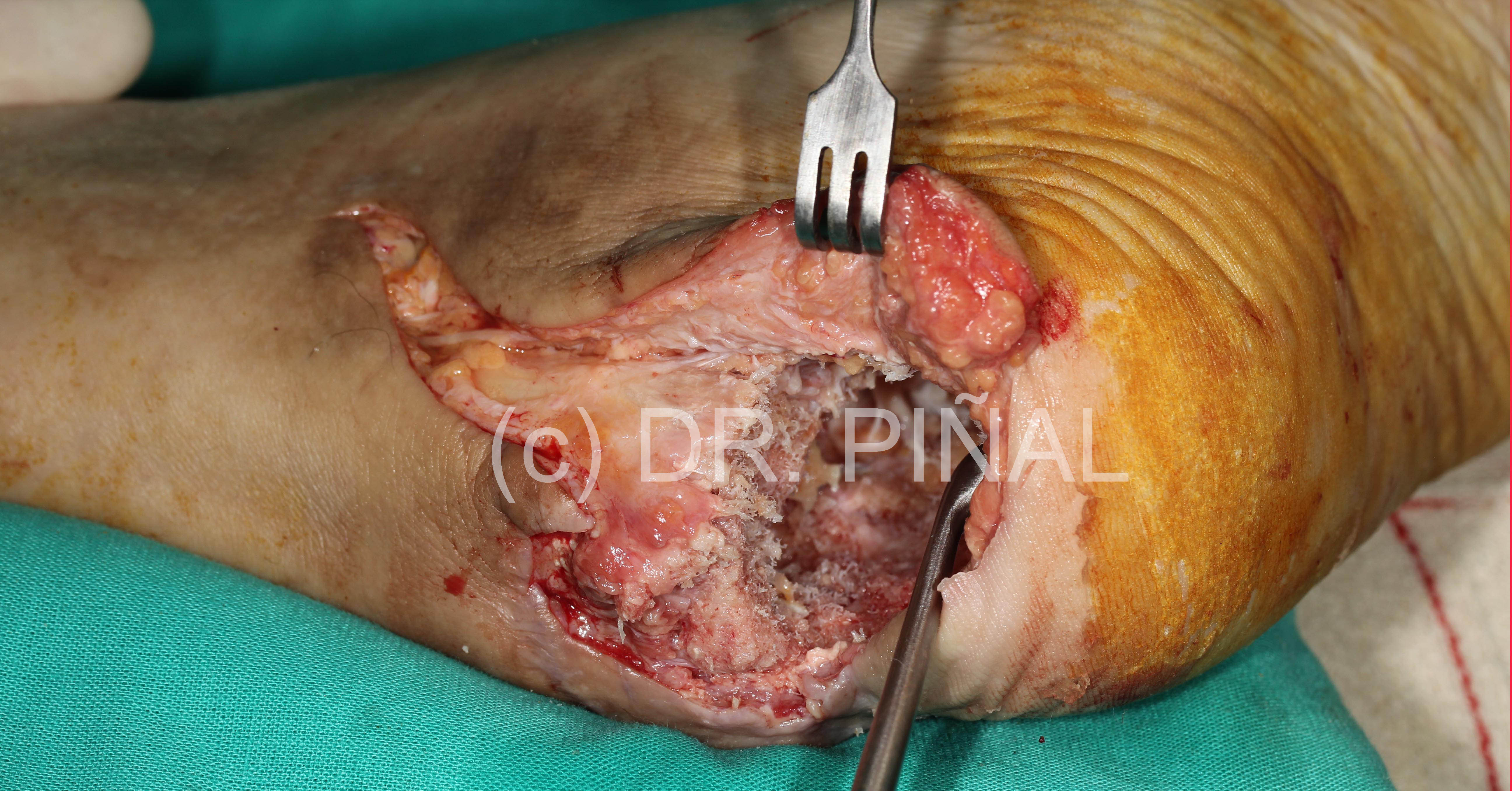
Antibiotic balls are introduced in the space opened, which remains 72 hours without coverage to facilitate the anti-infectious action.
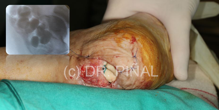
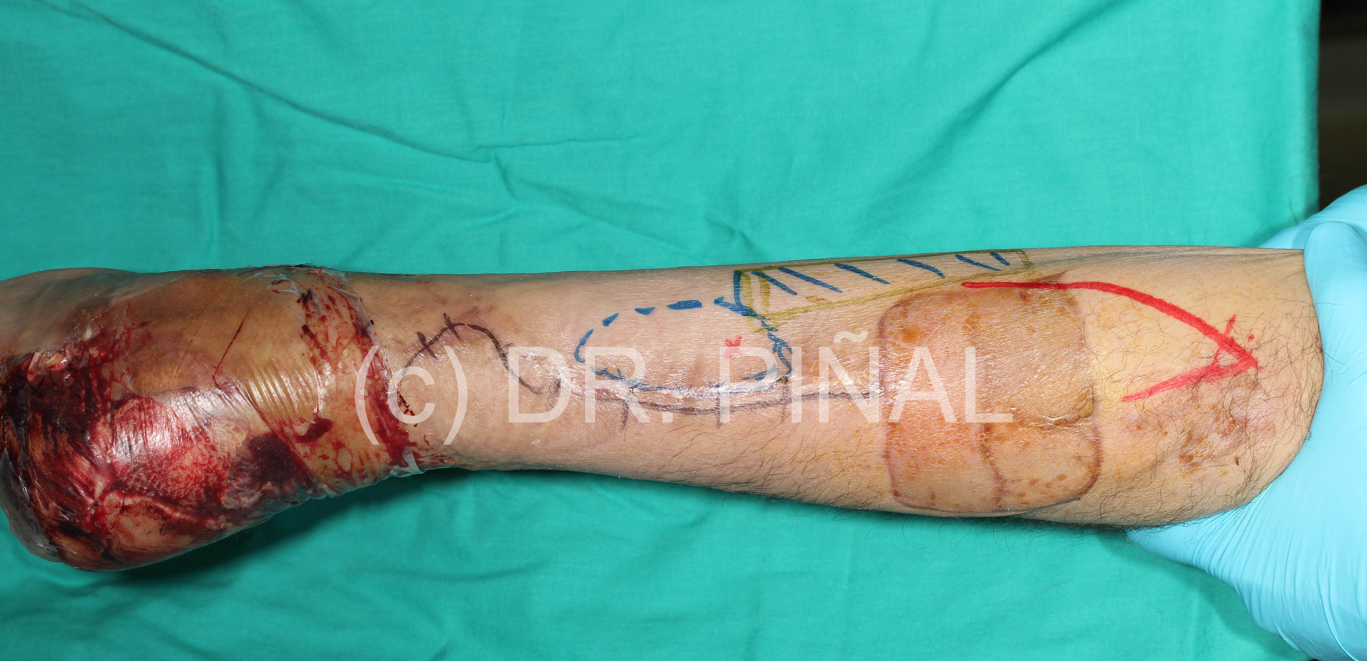
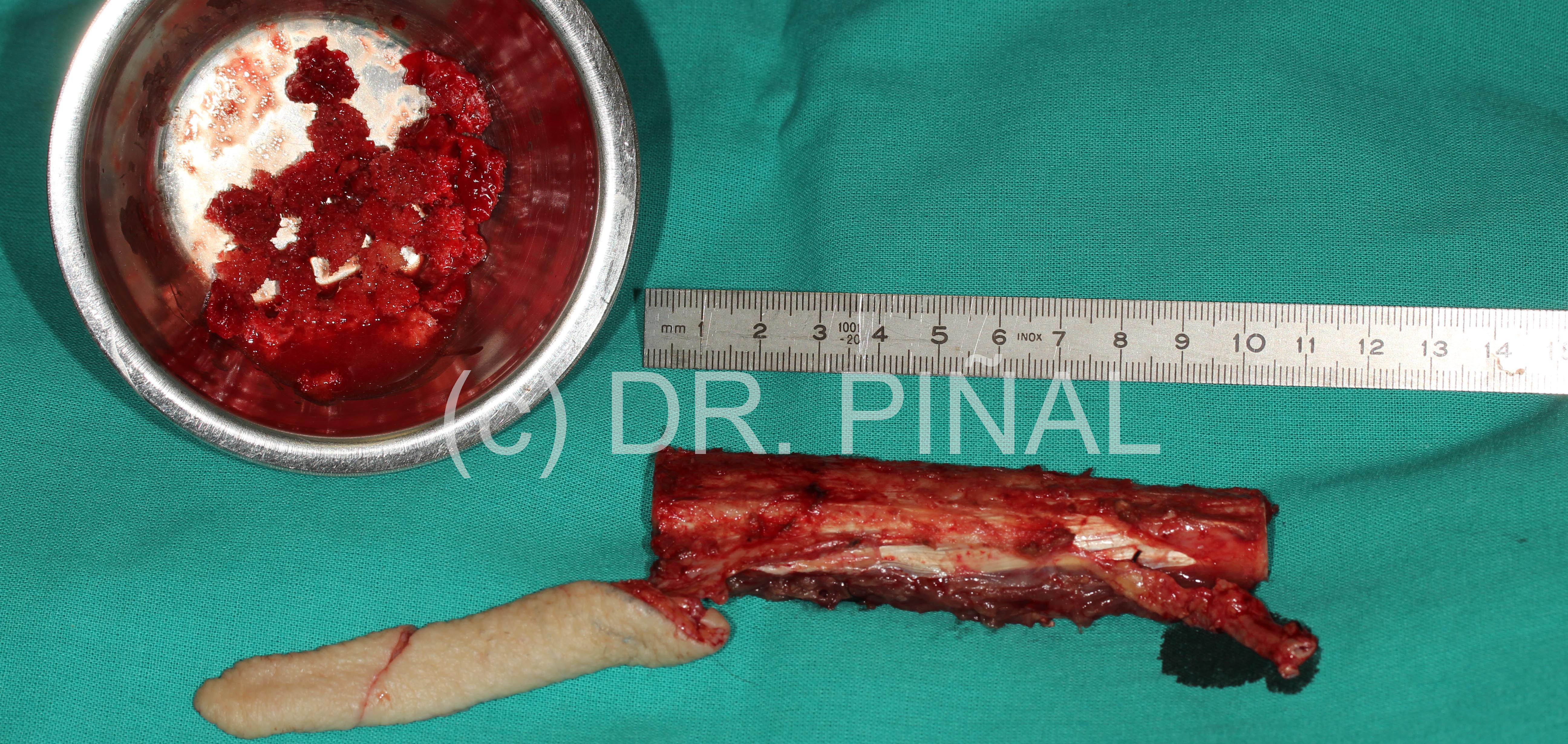
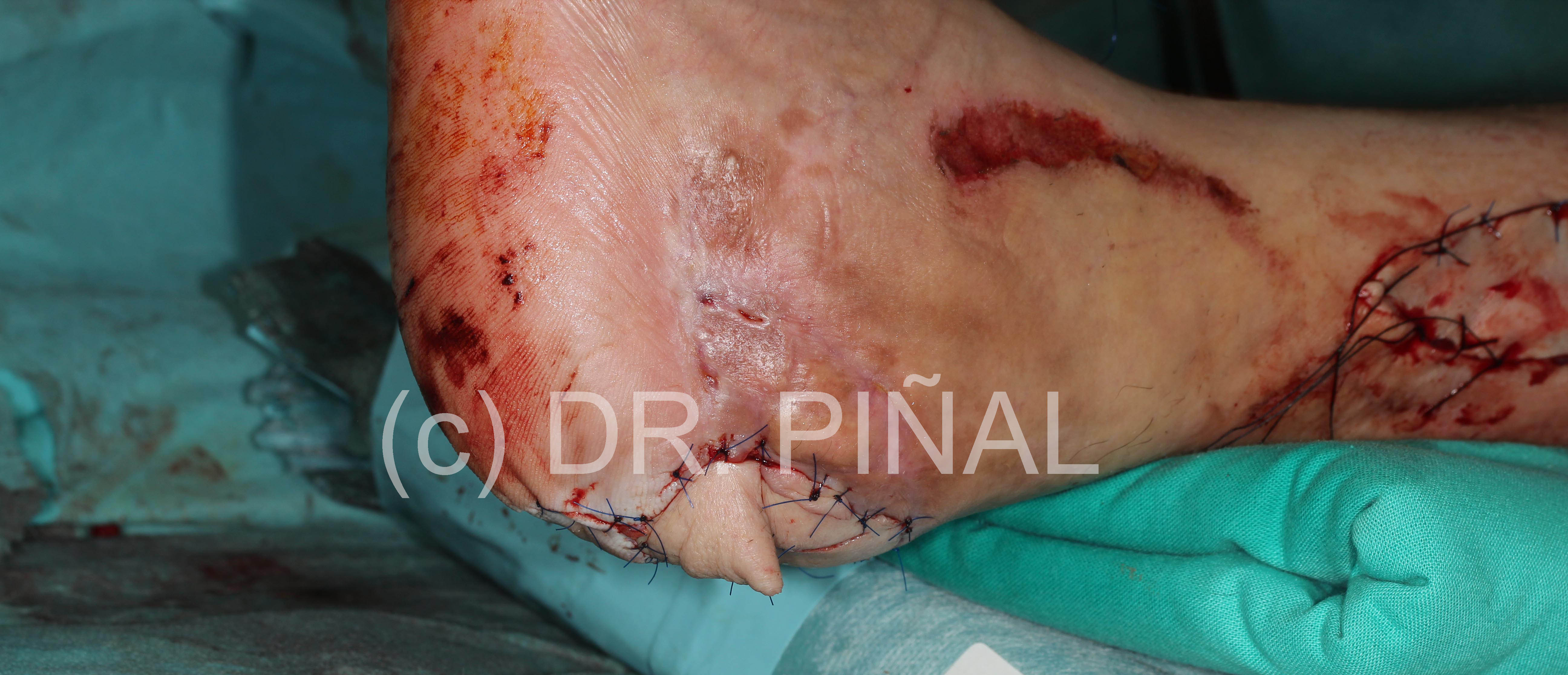
In a second step the operation area has to be provided with structure and blood supply (vascularization), for which a piece of vascularized fibula is grafted, with an autologous skin coverage and the use of spongy bone material from the patient as a filler.
The outcome
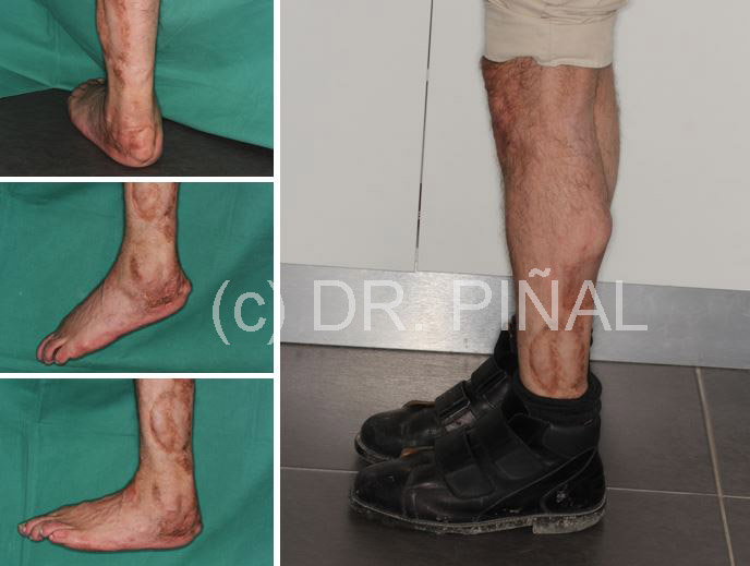
After the intervention of Dr. Del Piñal, the area is free from infection and the patient regains a level of functionality that allows him to return to work.
 es
es en
en fr
fr it
it ru
ru zh-hans
