然后,您可以通过手动翻译阅读此内容的相应英文翻译。 您也可以通过单击右上角的相应符号来查看西班牙语的原始文本。 通过此链接,您可以访问由Google翻译自动翻译的中文版本: https://bit.ly/3wiOHGV
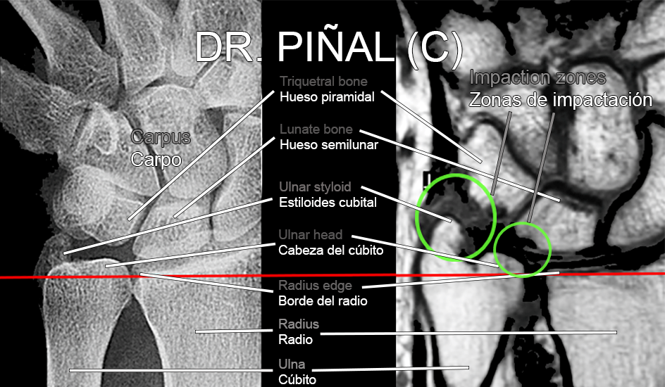
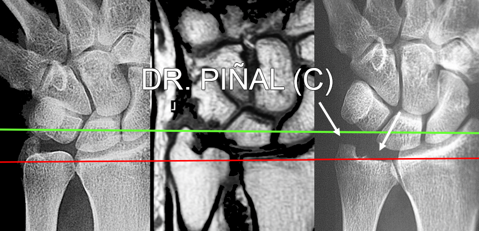
The patient, 43, suffers from a combined ulnar impaction syndrome in his right wrist caused by the excess length of the ulna and ulnar styloid, which collide with the carpal area. More specifically, the head of the ulna surpasses the edge of the radius and impacts against the lunate bone and the styloid, bigger than normal, does so against the triquetrum.

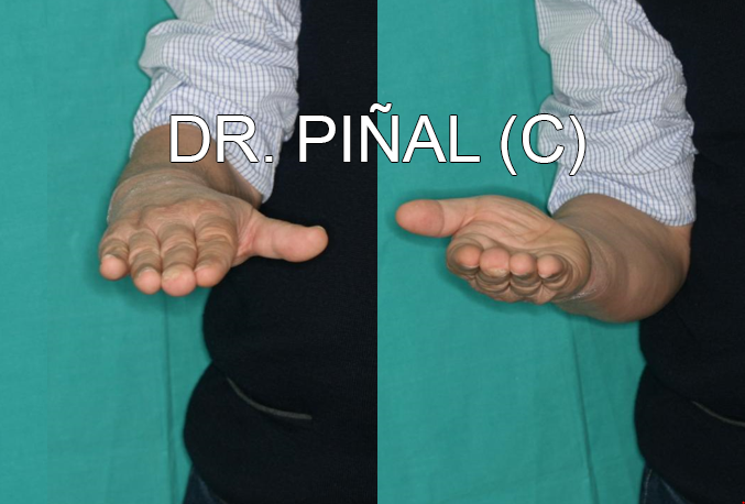
At the wrist, the ulna articulates with the radius – the so-called distal radioulnar joint – allowing hand and wrist rotation, that is, the movements of pronation (hand with the dorsum up) and its opposite, supination. In turn, it plays a prominent role as a load-bearing structure in these movements.
Positive ulnar variance or ulna plus, that’s to say, the ulna is longer than the radius at its distal ends, occurs in about 20% of the adult population. However, this characteristic becomes pathological, as in the case at hand, when the ulna repeatedly impacts on the ulnar carpal area.
The problem
Impaction syndrome causes pain in the patient on the ulnar side of the hand-wrist, as well as certain function problems. This degenerative pathology progressively degrades the triangular fibrocartilage and provokes bone damage to the lunate bone, due to its continuous collision with the head of the ulna and, in this case, also in the pyramidal one due to impacts with an abnormally sized ulnar styloid. Altogether, we find one of the pictures with several simultaneous injuries, common in a very complex area from an anatomical perspective.
Arthroscopy allows to visualize the degradation of the triangular fibrocartilage and the bone damage derived from the position of the head of the ulna and the ulnar styloid.
The clinical aims of Dr Piñal and his team focus on the elimination of ulnar pain and, by extension, the recovery of normal function in the affected limb.
The plan
Dr Piñal designs a procedure, based on his dry arthroscopy technique, to resect (cut) the ulna segment that exceeds the edge of the radius and the ulnar styloid fragment that impacts against the pyramidal bone, thus eliminating the two collision points.


The results
After surgery, the patient’s ulnar pain disappears and a complete recovery of movement is achieved.
患者证言:
 es
es en
en fr
fr it
it ru
ru zh-hans