 es
es en
en fr
fr it
it ru
ru ar
ar zh-hans
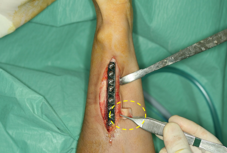
The patient, a nurse by profession, suffered a radius fracture in childhood that slowed the normal growth of this bone. This shorter radius length leads to an ulna plus situation, in which the head of the ulna is well above the radial border and impacts extensively (impaction syndrome) with the ulnar carpal area. This bone collision causes pain in the rotational movements of the distal radioulnar joint, that is, of the hand and wrist

From an anatomical perspective, we approach a particularly complex area, in which multiple elements converge in a very limited space (just over 1 cubit centimeter). For this reason, associated lesions are common, and if all aren’t treated pain and limitation will persist. In this case, the original fracture also caused a pseudoarthrosis of the ulnar styloid and the detachment of the triangular fibrocartilage (TFC) from the fovea of the ulna.
The TFC or articular disc is part of the distal radioulnar joint. It is a structure composed by a combination of fibrous and cartilaginous tissues, which is integrated -in turn- into the triangular fibrocartilage complex (TFCC), which allows us to perform harmonic movements and strength in the grip.
The problem
The patient presents a picture of pain in the pronosupination movements, caused by bone imbalances due to the fracture of the radius referred to in the presentation of the case.
The goals
The main clinical target is the anatomical correction of the position of the ulna and, consequently, the elimination of ulnar pain in the patient.
The plan
Dr Piñal designs a corrective procedure in one surgical stage, but treating several associated problems. First, the ulna is shortened so it recovers its normal anatomical position, aligned with the distal end of the radius. After removing the bone segment, the fragments are joined using an osteosynthesis plate.


After that, the fractured ulnar styloid is removed and a surgical anchor is used to act as a fixation for the reattachment of the TFC into the ulna fovea.


To suture the fibrocartilage, Dr Piñal uses all-inside suturing, that is, his arthroscopic suturing technique from inside the joint itself without additional incisions, described in his article ‘A technique for arthroscopic all-inside suturing in the wrist ‘(Journal of Hand Surgery European Volume (2010) 35: 475-479).
The results
The procedure eliminates ulnar pain and the distal radioulnar joint is in optimal function.
Postoperative status of the patient at four years, with adequate function and without pain.
Related content: